what does an abnormal s wave indicate S Wave Learn the Heart Healio
As the American Heart Association AHA describes it the right and left atria the upper chambers or ventricles make a wave called a P wave the bottom right and left chambers School of Health Sciences Practice Learning Cardiology Teaching Package A Beginners Guide to Normal Heart Function Sinus Rhythm Common Cardiac Arrhythmias The S Wave You will also have seen a small negative wave following the large R wave This is known as an S wave and represents depolarisation in the Purkinje fibres
what does an abnormal s wave indicate
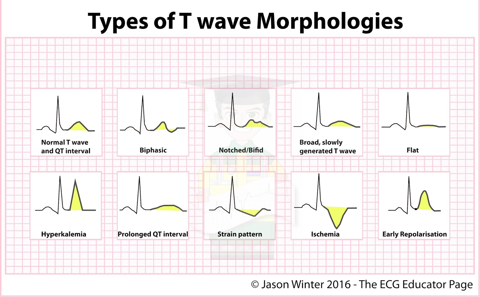
what does an abnormal s wave indicate
https://img.grepmed.com/uploads/552/morphologies-ecgeducator-medstudent-cardiology-twave-original.jpeg

Abnormal Ecg Chart
https://www.thecardiologyadvisor.com/wp-content/uploads/sites/17/2021/05/ECG_G_860096724.jpg
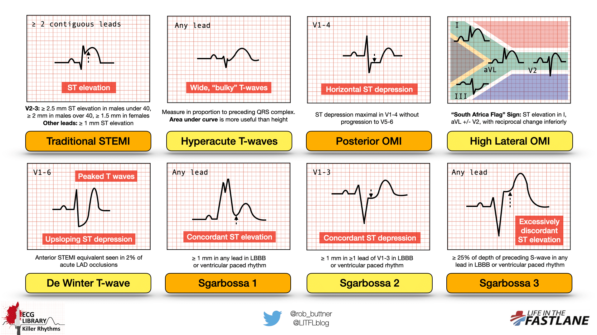
Fillable Online Ekg Library Litfl Ecg Library Basicshow To Read An Ecg
https://litfl.com/wp-content/uploads/2022/04/Killer-ECG-Part-2-Occlusion.png
Results Seeking help Medical treatments An abnormal EKG can sometimes happen due to a variation in the rhythm of your heart But it can also be an indicator of a more serious condition such Sinus arrhythmia is a kind of arrhythmia abnormal heart rhythm For the most common type of sinus arrhythmia the time between heartbeats can be slightly shorter or longer depending on whether you re breathing in or out Your heart rate increases when you breathe in and slows down when you breathe out
Your sinus rhythm is the pattern of electrical pulses from your sinus node your heart s pacemaker It s not uncommon for sinus rhythm to be too slow or too fast but Increased R wave amplitude and duration i e a pathologic R wave is a mirror image of a pathologic Q R S ratio in V1 or V2 1 i e prominent anterior forces Hyperacute ST T wave changes i e ST depression and large inverted T waves in V1 3 Late normalization of ST T with symmetrical upright T waves in V1 3
More picture related to what does an abnormal s wave indicate

Three Hearts Showing A P Wave QRS Complex And A T Wave Nursing
https://i.pinimg.com/originals/d2/0e/53/d20e539a7a0af0107ab54b119f8110d7.png
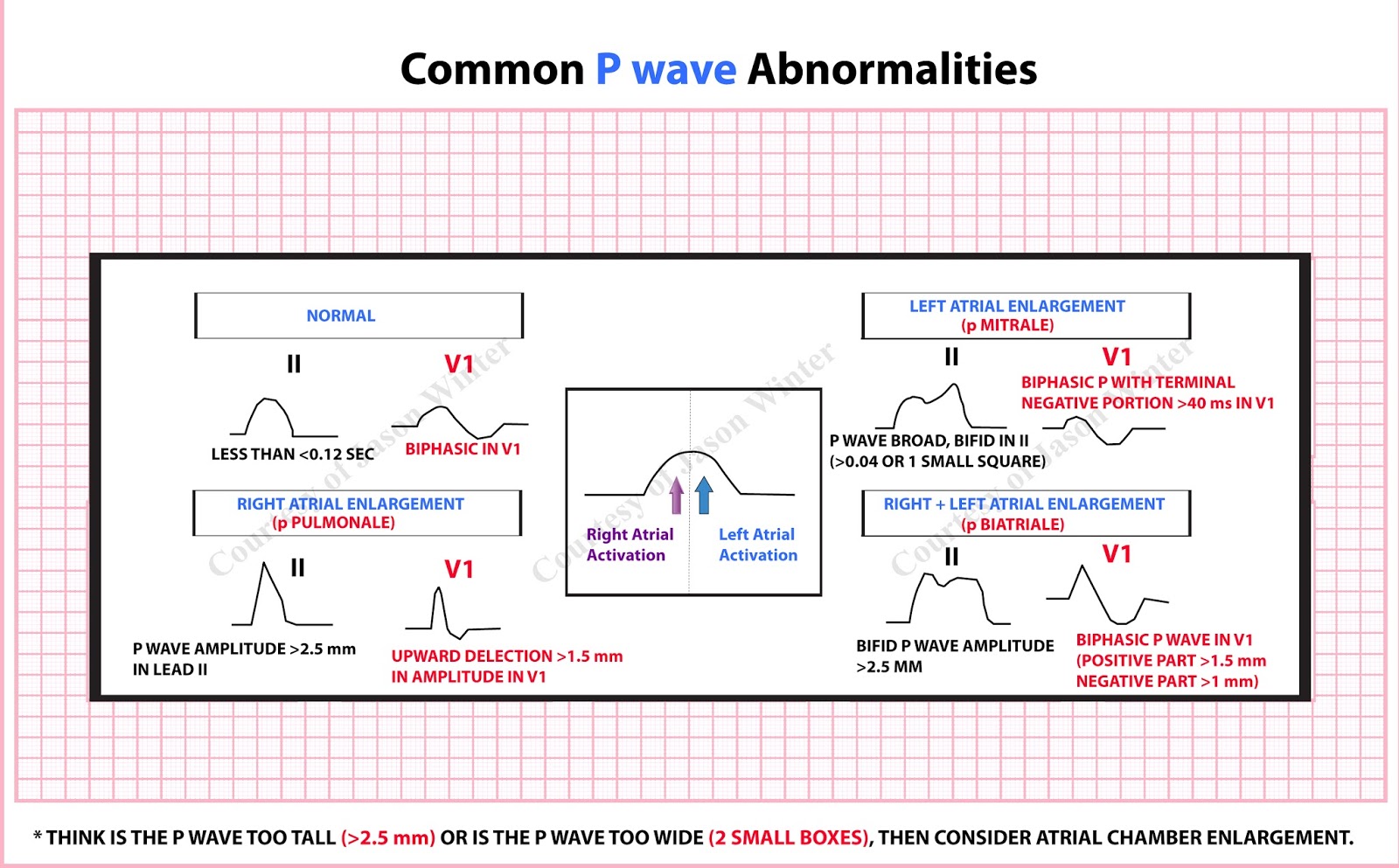
Common P Wave Abnormalities Diagnosis Cardiology GrepMed
https://img.grepmed.com/uploads/519/atrialenlargement-enlargement-ecgeducator-medstudent-cardiology-original.jpeg
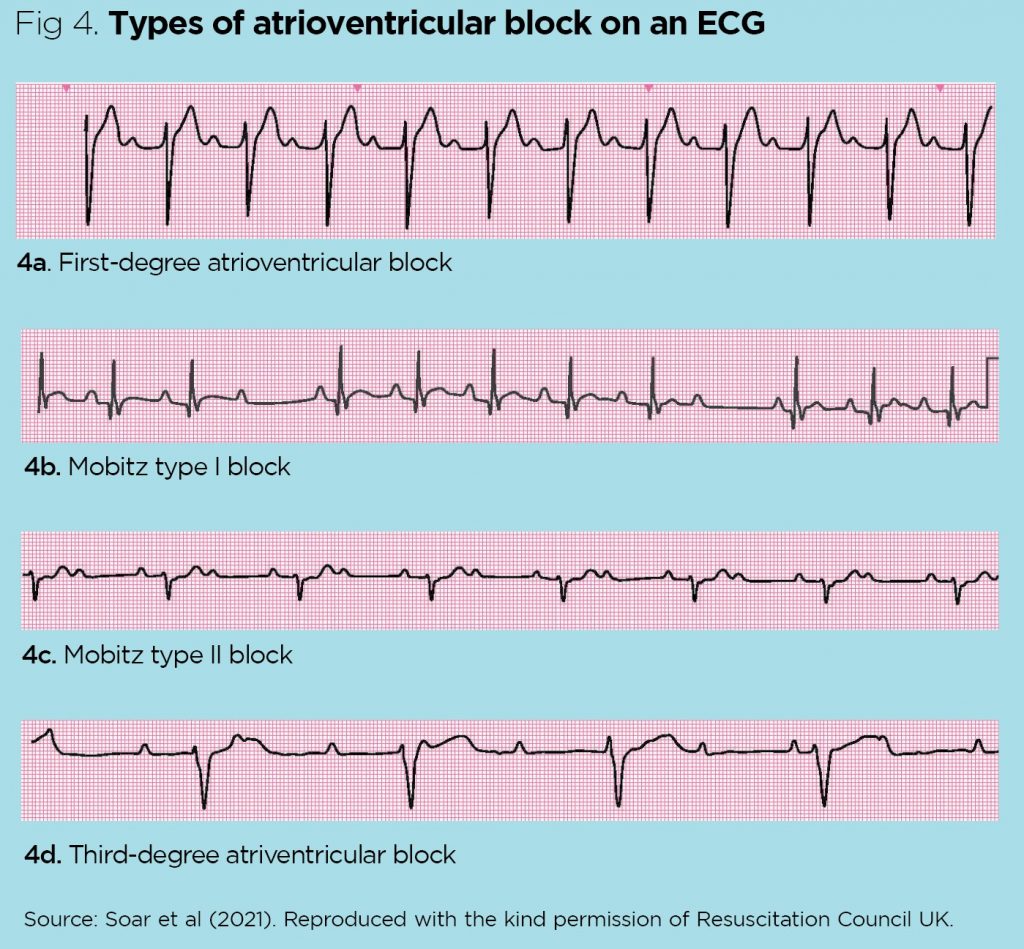
Different Types Of Abnormal Ekg
https://cdn.ps.emap.com/wp-content/uploads/sites/3/2021/07/Fig-4.-Types-of-atrioventricular-block-on-an-ECG1-1024x949.jpg
Unlike early repolarization T waves are usually low amplitude and heart rate is usually increased May see PR segment depression a manifestation of atrial injury Other Causes Left ventricular hypertrophy in right precordial leads with large S waves Left bundle branch block in right precordial leads with large S waves Advanced The two R waves indicate the depolarisation of the right and left sides of the heart at different times the right depolarises after the left You can remember the pattern with the word M arro W there is M in V1 and
Left bundle branch block produces a dominant S wave in V1 with broad notched R waves and absent Q waves in the lateral leads Hyperkalaemia is associated with a range of abnormalities including peaked T waves Tricyclic poisoning is associated with sinus tachycardia and tall R wave in aVR Treatments Uses Summary Sometimes an abnormal EKG reading is a normal variation in a person s heart rhythm In other cases it may be due to an underlying condition of the heart or a
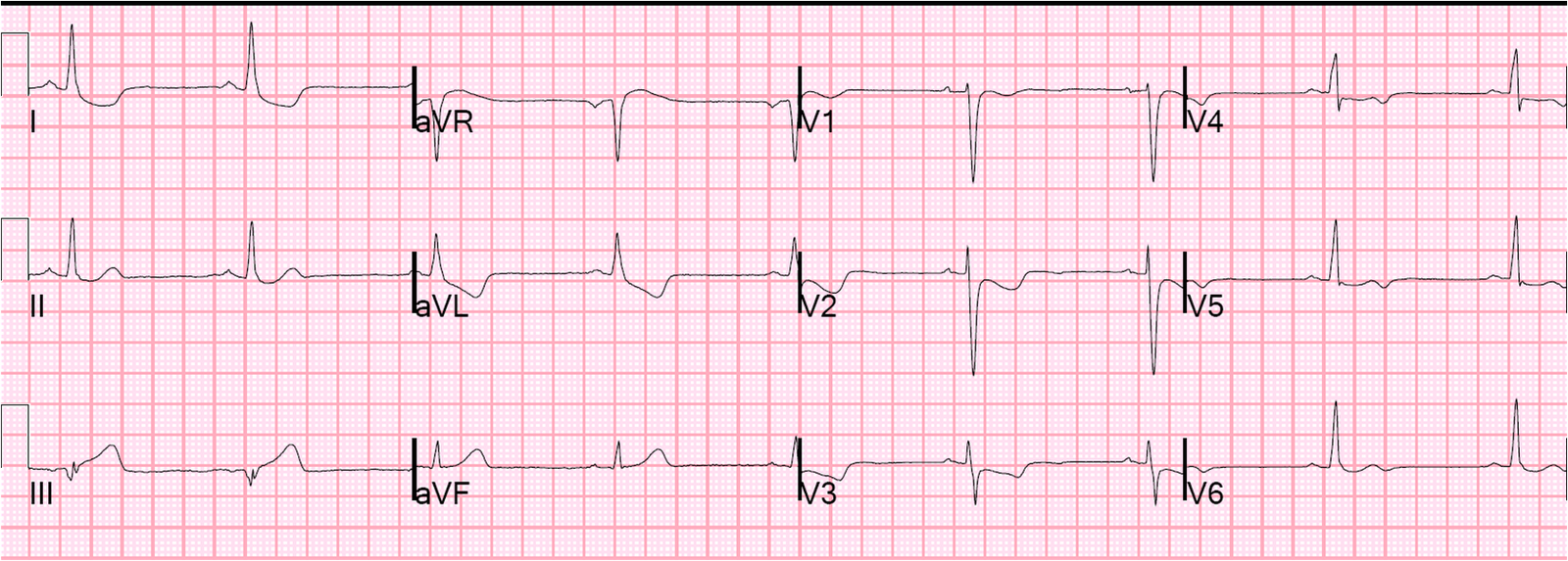
Dr Smith s ECG Blog A Man In His 20 s With Syncope What Is Diagnosis
http://4.bp.blogspot.com/-fDw-yqv_Dng/UHw-dNAoF5I/AAAAAAAABsY/SIcedDNODjE/s1600/ECG+1+1920.png
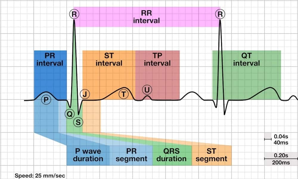
R Wave LITFL ECG Library Basics
https://litfl.com/wp-content/uploads/2018/10/ECG-waves-segments-and-intervals-LITFL-ECG-library-3.jpg
what does an abnormal s wave indicate - An abnormal p wave because the excitation has begun somewhere away from the SA node Normal QRS Normal beats after the abnormal one Junctional escape No p waves Normal QRS Slightly slower rate 75bpm max Ventricular escape